Excision of incarcerated, gangrenous small bowel in a patient with septic shock
A 49 yr old lady presented to the hospital with a 4 day history of abdominal pain. She was a patient with a high BMI of about 35, known to have Diabetes and hypertension. She was diagnosed to have an incarcerated incisional hernia. Echo at the time of admission showed hypokinesia of the inferior wall. She had a fair LV function with an EF of 58%.
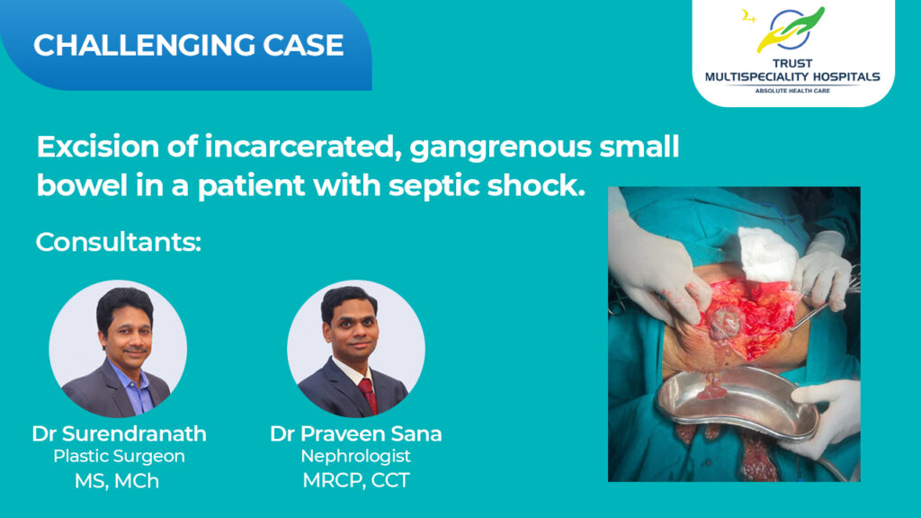
She underwent an emergency laparotomy and incarcerated*, gangrenous** small bowel was found in an incisional hernia. Gangrenous bowel was excised and end to end anastomosis was done by the surgical team. She developed septic shock post op and required high ionotropic support. She developed oliguric AKI with metabolic acidosis. In view of circulatory shock, she was provided CRRT*** support in ICU for 36 hrs by the Nephrology and intensive care teams. She made a good recovery and renal function normalized. She was discharged in a stable condition.
*Incarcerated hernia is when a part of the bowel protrudes out of the abdomen and cannot be pushed inside.
**gangrene is the death of a body tissue due to lack of Oxygen suppy
*** CRRT is a 24 hour non-stop dialysis done in patients who are extremely unwell and have kidney failure.
Catheter direct thrombolysis of occluded SMA
A 45 yr old male patient was diagnosed with peripheral vascular disease in 2017 when he had presented with claudication pain in the left lower limb. He was a chronic smoker. CT angio at that time revealed diffuse atherosclerotic disease with significant narrowing and non opacifation of right internal iliac, bilateral femoral, popliteal and tibial arteries. He was treated with Aspirin, Clopidogrel, Heparin, Cilostazol and Pentoxifylline. He was later given 6 cycles of PGE-1 therapy with which he had partial improvement. Since late 2019, he was poorly compliant to medication with irregular follow up with recurrence of claudication pain.
He presented now with a one week history of left sided abdominal pain, predominantly after food intake associated with black stools. A possibility of mesenteric ischemia was considered and USG abdomen with Doppler revealed thickened jejunal folds with SMA thrombosis. CT angiogram also revealed SMA thrombosis with circumferential thickening of jejunum with associated fat stranding. He was anticoagulated with UFH while monitoring APTT and was later changed to LMWH. He was monitored for any evidence of bowel gangrene. He continued to have pain abdomen following food intake. Doppler after 5days and 10 days of heparin therapy revealed non clearance of thrombus with no blood flow. Then we took the help of our esteemed and very experienced cardiology colleagues.
Patient was taken up for SMA intervention. Occlusion of SMA was confirmed via angiography (1). SMA was crossed with a pilot wire and dilated with 4 mm balloon. Flow not established despite dilatation. In view of persistent thrombosis, reperfusion Catheter was placed in SMA and Streptokinase was infused over 48 hrs at 1ml / hr resulting in good reperfusion (2).